
How 3D/4D Ultrasound Technology Enhances Prenatal Care - Samad Hospital
A closer look at advanced pregnancy imaging and its benefits for mothers and babies
Ultrasound scans are one of the most reassuring and exciting parts of pregnancy, allowing parents to see their growing baby while helping doctors monitor the baby’s development and health. Over the years, ultrasound technology has evolved significantly. Beyond the traditional 2D scans, modern medicine now offers advanced 3D and 4D ultrasound imaging, providing clearer, more detailed, and dynamic views of the unborn baby. These advancements not only create emotional bonding moments for parents but also play a crucial role in enhancing prenatal care, early diagnosis, and treatment planning.
Understanding 3D and 4D Ultrasound Scans
A 2D ultrasound uses sound waves to create flat, black-and-white images of the baby and internal organs. While extremely useful medically, the images are sometimes harder for parents to interpret.
A 3D ultrasound goes one step further by capturing multiple two-dimensional images from different angles and combining them to form a realistic three-dimensional image. This allows clearer visualization of the baby’s facial features, limbs, body shape, and certain anatomical structures.
A 4D ultrasound adds time to 3D imaging. This means parents and doctors can see real-time movements—such as the baby yawning, blinking, stretching, smiling, or sucking its thumb. It feels like watching a short video of the baby inside the womb.
How These Advanced Scans Improve Prenatal Care
While many parents consider 3D and 4D scans primarily as exciting bonding experiences, they actually provide significant medical benefits. These technologies allow doctors to see clearer, sharper details and identify issues that might not be easily visible in standard scans.
Better Visualisation of Fetal Development
One of the most important benefits is enhanced clarity. With 3D imaging, doctors can view precise structures of the baby’s face, skull, spine, limbs, and organs. This allows for:
-
Detailed observation of fetal development
-
Clearer tracking of growth milestones
-
Improved assessment of movements and physical activities
This high level of detail offers vital insight into the baby’s wellbeing.
Early Detection of Congenital Abnormalities
3D and 4D ultrasounds are especially helpful in identifying certain birth defects earlier and more accurately. These include:
-
Cleft lip or palate
-
Facial deformities
-
Spine abnormalities like spina bifida
-
Limb deformities
-
Some heart and abdominal wall abnormalities
Early detection allows doctors to plan appropriate care during pregnancy, arrange additional monitoring, and prepare necessary treatment soon after birth if required. For parents, early knowledge also provides time to prepare emotionally and practically.
Improved Assessment of Placenta and Amniotic Fluid
Healthy placenta function and proper amniotic fluid levels are essential for the baby’s nourishment and safety. Advanced ultrasound imaging helps evaluate:
-
Placental position and health
-
Blood flow patterns
-
Amniotic fluid volume
-
Possible risk conditions such as placenta previa
Timely identification of such conditions helps doctors take preventive measures, reducing risks to the mother and baby.
Better Monitoring in High-Risk Pregnancies
3D and 4D ultrasounds are particularly beneficial for women experiencing high-risk pregnancies, including:
-
Mothers with diabetes or hypertension
-
Women with a history of pregnancy complications
-
Multiple pregnancies (twins or more)
-
Advanced maternal age
-
Previous congenital abnormalities
For these pregnancies, continuous and accurate monitoring is crucial. These advanced scans provide clearer insight, helping specialists closely watch the baby’s growth and respond quickly if concerns arise.
Strengthening the Emotional Bond Between Parents and Baby
Beyond medical advantages, 3D and 4D ultrasounds create a deeply emotional experience. Unlike 2D images that are often difficult to interpret, these advanced scans show a lifelike view of the baby, helping parents:
-
See their baby’s facial expressions
-
Watch movements and gestures
-
Feel more connected to the pregnancy
-
Reduce anxiety by visually confirming the baby’s wellbeing
For many families, these moments become cherished memories and contribute positively to emotional health during pregnancy.
Is 3D/4D Ultrasound Safe?
Safety is always a primary concern for expectant parents. 3D and 4D ultrasounds use the same sound-wave technology as traditional scans and do not involve radiation. When performed by trained professionals and used appropriately for medical purposes, they are considered safe and reliable.
Doctors usually recommend performing these scans at certain stages for best clarity and diagnostic value, often between 24 to 34 weeks of pregnancy, depending on medical needs.
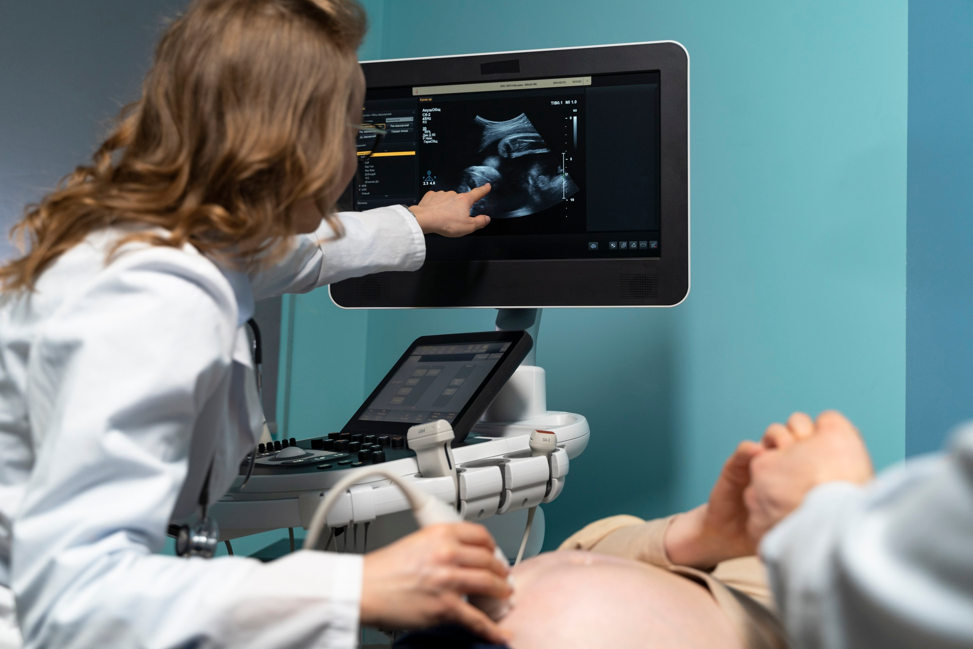
What to Expect During the Scan
The procedure is generally simple, comfortable, and similar to a regular ultrasound.
-
A gel is applied to the abdomen
-
A handheld device (transducer) moves across the belly
-
Images appear on a monitor in real time
The duration may vary depending on fetal position and medical evaluation requirements. Sometimes, if the baby is not in a suitable position, the doctor may ask the mother to change posture or return at another time to get clearer images.
When is 3D/4D Ultrasound Recommended?
Doctors may suggest these scans if:
-
Detailed assessment of fetal anatomy is required
-
A suspected abnormality needs clearer evaluation
-
High-risk monitoring is necessary
-
Parents desire advanced bonding imaging along with clinical review
However, these scans should always be done at reputable medical centres under expert supervision.
Conclusion
3D and 4D ultrasound technology represents a significant advancement in prenatal care. By offering clearer, more detailed images, it enhances diagnosis, supports early treatment planning, improves monitoring in high-risk pregnancies, and strengthens emotional bonding between parents and their unborn baby. Beyond the joy of seeing a baby’s face and movements, these scans play a powerful role in ensuring safer pregnancies, healthier outcomes, and greater peace of mind for expectant families.
Leave a Comment