
Growth Scan During Pregnancy: Baby's Development
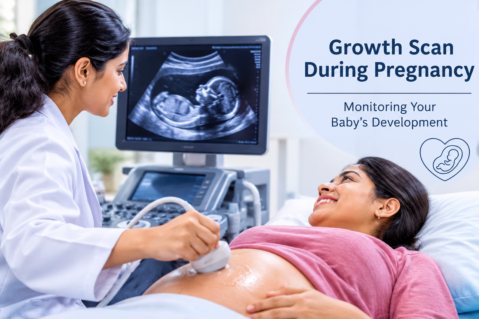
Carrying a child for nine months means adjusting to physical changes, riding a range of emotions and passing through several important medical checkpoints. One of the most useful things doctors can do along the way is follow how the baby is growing, since it shows how healthy the fetus is and brings possible problems to light early. Standard antenatal appointments tell the team mostly about the mother. Special ultrasound tests open a window onto the baby itself, and the growth scan is a prime example.
In this ultrasound, the doctor looks at how large the baby has become, roughly what it weighs, the way it lies in the womb and how its body is developing as a whole. From the findings, the team can tell whether growth is following the normal pattern or whether the pregnancy needs a closer watch. Parents-to-be who want to know why the scan matters can consult a Fetal Medicine Specialist in Kerala, who can tailor advice to their situation.
What Is a Growth Scan During Pregnancy?
Think of a growth scan as a later-stage ultrasound dedicated to checking how the baby’s body is coming along. Various body parts are measured, and the readings are combined to estimate how much the baby weighs.
Its role differs from the first scan in early pregnancy, which mainly confirms the pregnancy and dates it. The growth scan is about size and progress further along.
During the exam, the doctor typically records:
- The size of the baby’s head, measured around its edge.
- The measurement around the belly.
- The length of the thigh bone.
- A calculated weight estimate.
- How much amniotic fluid surrounds the baby.
- The placenta’s location and how it looks on screen.
- The baby’s orientation in the uterus.
Each reading is then compared with what is typical for the same week of pregnancy.
Why Is a Growth Scan Important During Pregnancy?
No two babies grow at exactly the same pace. Even so, checking at intervals shows doctors whether a particular baby is on course. A scan can uncover things that feeling and measuring the abdomen from outside would not.
1. Monitoring Fetal Growth
Tracking size and progress is the scan’s central job. Doctors take the head, belly and thigh-bone readings and turn them into a weight estimate, which also reveals how growth is trending. If the numbers fall noticeably short of the norm or run well ahead of it, the doctor might order more investigations or book another scan.
2. Identifying Fetal Growth Restriction
A baby that is not gaining size at the expected pace is said to have fetal growth restriction (FGR). The reasons can include placental trouble, a health condition in the mother or other pregnancy-related factors. The scan lets doctors spot babies who measure small for their stage. Follow-up often involves Doppler tests and closer observation to assess blood flow and how the baby is faring.
3. Assessing Amniotic Fluid Levels
The fluid that bathes the baby shields it from knocks and gives it room to move and mature. Levels that run unusually low or high can need treatment, depending on the cause and the point reached in the pregnancy.
4. Evaluating Placental Health
Everything the baby receives in oxygen and nourishment passes through the placenta, so how it lies and looks on ultrasound is informative. Where there is reason for concern, doctors can arrange extra tests of its performance and blood supply.
5. Supporting Pregnancy Management
What the scan shows helps the doctor choose between continuing standard antenatal care and stepping up surveillance. That decision matters most when the mother has a medical condition or a problem has already come up during the pregnancy.
When Is a Growth Scan Recommended?
Scheduling is individual. It turns on the mother’s health history, the course of the pregnancy and the doctor’s own assessment.
A typical growth check takes place in the last trimester, usually somewhere between weeks 28 and 36, but there is no single timetable for everyone. If a concern about development arises, the scan may be brought forward or repeated.
Close growth monitoring is particularly worthwhile when:
- A previous pregnancy involved restricted growth in the baby.
- The mother’s blood pressure is high.
- She has developed diabetes during pregnancy.
- She is expecting more than one baby.
- A routine visit raises a question about the baby’s size.
- She notices the baby moving less than usual.
- The placenta has certain irregularities.
- She has a health condition that could influence growth.
Fitting the schedule to the person ensures the baby is followed adequately, without scans that add nothing.
How Is a Growth Scan Performed?
Nothing invasive is involved. The scan uses high-frequency sound waves that are turned into pictures of the baby. The mother rests on an examination couch, gel goes onto her abdomen, and the sonographer or doctor passes a probe over the skin.
The scan can include the following stages:
Step 1: Fetal Position Assessment
The doctor first notes how the baby is lying so that the readings come out accurate.
Step 2: Biometric Measurements
The head, abdomen and thigh bone are measured, and those figures feed the calculation of weight.
Step 3: Amniotic Fluid Assessment
Ultrasound is used to gauge how much fluid lies around the baby.
Step 4: Placental Evaluation
When there is a medical reason, the position and appearance of the placenta are checked.
Step 5: Doppler Assessment
If necessary, a Doppler study follows blood moving through chosen vessels in the baby and the placenta.
The time taken varies with the baby’s position and how thorough the examination needs to be. For most mothers it is a comfortable experience.
Understanding the Measurements in a Growth Scan Report
A scan report is full of numbers. When parents understand what each one stands for, appointments become much easier to follow.
Head Circumference (HC)
This is the baby’s head measured around its edge. It shows how the head is developing and is one input into the weight estimate.
Abdominal Circumference (AC)
The abdomen is measured all the way around. The result is among the most important growth indicators because it tends to mirror how well the baby is being fed and how it is developing overall.
Femur Length (FL)
This refers to the thigh bone. It reflects the growth of the skeleton, and the weight estimate draws on it together with the other readings.
Estimated Fetal Weight (EFW)
A formula that combines several scan readings gives this approximate weight for the day of the scan. It remains an approximation, so a little variation is normal, and the doctor reads the figure alongside the baby’s age in weeks and its growth history.
What Is Doppler Ultrasound and Why Is It Used?
Doppler is an ultrasound variation that studies how blood flows in particular vessels. Doctors turn to it for extra information on how well the placenta is circulating blood and how the baby is coping.
Depending on the situation, blood flow may be checked in:
- The umbilical artery.
- The middle cerebral artery.
- The uterine arteries.
- Further vessels of the baby, where relevant.
If the flow looks abnormal, the baby may need to be watched more closely. The doctor reads Doppler results together with growth readings, the mother’s health and other details.
Whether a Doppler test is needed, and what the results imply for the pregnancy, can be explained by a Fetal Medicine Specialist in Kerala.
Factors That May Affect Baby’s Growth During Pregnancy
Growth is shaped by things related to the mother, the baby and the placenta. The usual ones are:
Maternal Nutrition: A diet short on nutrients can hold back the baby’s growth, whereas balanced eating helps both mother and child.
High Blood Pressure: Raised pressure, including disorders that appear only in pregnancy, can restrict circulation through the placenta and influence how the baby develops.
Gestational Diabetes: If blood sugar is not well managed, a baby may grow larger than normal and other complications may arise.
Placental Problems: A placenta that is not working properly can deliver less oxygen and fewer nutrients to the baby.
Multiple Pregnancy: Twins or more call for additional growth checks, as each baby can develop at its own rate.
Lifestyle Factors: Smoking, drinking and contact with some substances can harm the baby’s development.
Finding and managing these issues with medical supervision leads to better pregnancy care.
What Happens If the Baby Is Not Growing as Expected?
A result outside the expected range does not by itself mean something is seriously wrong. Ultrasound readings have limitations, so doctors take several things into account first.
Where growth is a concern, the care team may propose:
- Scans repeated after a suitable interval.
- Doppler studies.
- More careful checking of the amniotic fluid.
- A look at the mother’s blood pressure and other health matters.
- Further tests of the baby’s wellbeing.
- A personal plan for when and how the baby is born.
The course taken depends on the baby’s growth pattern, how far along the pregnancy is, the Doppler findings and the mother’s overall health. Parents should avoid drawing conclusions from a single number and talk to their doctor instead.
How Can Expectant Mothers Support Healthy Fetal Growth?
Not every growth problem can be prevented, but sensible habits give a pregnancy the best footing.
Follow a Balanced Diet: Build daily meals around fruit, vegetables, whole grains, protein and other nourishing foods.
Attend Regular Antenatal Check-ups: Keeping appointments lets doctors follow the mother’s health and pick up complications early.
Take Prescribed Supplements: Folic acid, iron, calcium and other supplements should be taken exactly as the doctor advises.
Stay Physically Active: Gentle exercise that the doctor has approved supports health through pregnancy.
Monitor Baby’s Movements: Learn how your baby normally moves, and call your doctor promptly if the pattern slows or shifts.
Manage Existing Medical Conditions: Diabetes, high blood pressure and similar problems need regular monitoring and treatment from the first weeks to the last.
Choosing the Right Fetal Medicine Care
Fetal medicine units concentrate on finding and following up concerns during pregnancy. They bring together advanced ultrasound, diagnosis before birth and care built around each patient.
Expectant mothers visiting Samad Hospitals can get expert advice on following their baby’s growth and on other worries about the pregnancy. A careful evaluation lets doctors see how the baby is developing and recommend more tests if required.
Meeting an experienced Fetal Medicine Specialist in Kerala can also help parents understand their scan results, discuss any risks and make decisions with greater assurance as the pregnancy advances.
Conclusion
As a way of following a baby’s development and judging key points of fetal health, the growth scan is hard to beat. By studying measurements, estimated weight, amniotic fluid and the placenta, doctors can recognise potential growth problems and plan the right care.
Because pregnancies differ, the need for a growth scan depends on the person. Attending antenatal visits, having investigations on time and keeping up healthy habits all strengthen monitoring.
For parents hoping for trustworthy pregnancy assessment and specialised fetal care, Samad Hospitals gives them the opportunity to share concerns and receive suitable medical guidance. Knowing about fetal growth and keeping to the recommended monitoring plan can leave families feeling informed and confident as the pregnancy nears its end.
Leave a Comment